Diabetic Eye Examinations
Diabetes is the number 1 cause of Adult Blindness in the UK
An eye examination can sometimes detect diabetes, high blood pressure or neurological abnormalities.
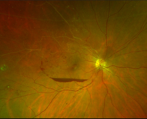
The markers for diabetic retinopathy are micro-aneurysms, haemorrhages and exudates. Diabetic retinopathy can be classified as mild, moderate and severe. When exudates, haemorrhages and micro-aneurysms associated with thickening within the foveal area then we term this as Diabetic maculopathy.
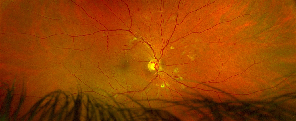
The National diabetic screening recommends that diabetics should have their pupils dilated and 2 images per eye taken with a retinal camera. We at Radlett opticians use a stricter criteria for diabetic eye examination using Optomap Ultra-widefield camera, OCT scanners and ERG to diagnose diabetic retinopathy early.
DAYTONA PLUS
Conventional cameras offer a max of 40% image of the retina.
Optomap Ultra-widefield retinal imaging offers:
-
200 degree or 80% of the retina.
-
Uninterrupted pathology of the peripheral retina.
-
Auto-fluorescence facility to monitor active metabolic changes of the retina.
-
View of early and subtle signs of diabetic retinopathy in an otherwise asymptomatic diabetic patient.
-
Unprecedented view of the complete retina without dilating the pupil.
OPTICAL COHERENCE TOMOGRAPHER
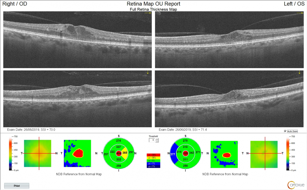
OCT allows a more accurate diagnosis of diabetic maculopathy.
Retinal imaging only allows a 2-dimensional view of the retina but unable to show retinal thickening.
OCT uses light technology to give a cross-sectional 3D view of the retina.
At Radlett opticians we use angio-OCT to locate the vessels causing micro-aneurysms, haemorrhages and exudates. OCT can pin-point retina / macula oedema normally missed by fundus photography screening.
ERG & VALEDA
Diopsys flicker ERG has been shown to provide important information regarding global retinal ischemia and the subsequent risk of developing new vessels called Proliferative diabetic retinopathy.
Results of the flicker ERG allows practitioners to administer intra-vitreous injections and help monitor the efficacy of the treatment on the proliferative retinopathy as well on the diabetic maculopathy.
Valeda photo-biomodulation is being trialled in Germany at present to treat diabetic maculopathy. It uses low level light therapy to retinal tissues to provide oxygen for the cells to function efficiently as well as anti-inflammatory benefits to the retinal vessels. For diabetic maculopathy the treatment is to stop the vascular leakage at the macula and help resolve the macula oedema.


